Bone Cross Section Diagram / Sketch And Label Of A Cross Section Of A Long Bone Long Bone Cross Section Worksheet Teaching Resources As The Names Suggest Compact Bone Looks Compact And The Spongy Bone / In a cross section of a bone we can see two types of bone tissue:
Bone Cross Section Diagram / Sketch And Label Of A Cross Section Of A Long Bone Long Bone Cross Section Worksheet Teaching Resources As The Names Suggest Compact Bone Looks Compact And The Spongy Bone / In a cross section of a bone we can see two types of bone tissue:. Diagram with articular cartilage, marrow, medullary cavity and periosteum. Spinal cord spinal column anatomy information myvmc. For example, to read this diagram literally, since the cartilage can be seen inside the cutaway section of bone, it. Explaned distal and proximal epiphysis. Haz tu selección entre imágenes premium sobre bone cross section de la más alta calidad.
Tetraplegia And Paraplegia Spinal Neural Disorder Medical Vector Illustration Diagram With Female Back Bone Cross Section Stock Vector Illustration Of Medicine Health 117454822 from thumbs.dreamstime.com Vector illustration scheme of bone cross section. Compact bone is the outer layer and the spongy bone forms the inner layer. As with other tools applied to petroleum development. Diagram with articular cartilage, marrow, medullary cavity and periosteum. Explaned distal and proximal epiphysis. This is why anatomical position for the hand in medical diagrams is with the thumb pointing out instead of the more natural pointing in, so the radius and ulna are parallel instead of crossing over each other. 850 x 1270 png 173kb. Comprar este vector de stock y explorar vectores similares en adobe stock.
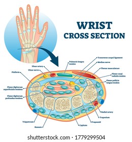
Cross section through middle metacarpal bones of vector.
This is why anatomical position for the hand in medical diagrams is with the thumb pointing out instead of the more natural pointing in, so the radius and ulna are parallel instead of crossing over each other. They build the entire picture, improve your understanding, consolidate the information and facilitate recall. Diagram with articular cartilage, marrow, medullary cavity and periosteum. Geological cross sections are graphical representations of vertical slices through the earth used to clarify or interpret geological relationships with or without accompanying maps. Jump to navigation jump to search. Cross section of a human bone. Vector illustration scheme of bone cross section. Diagram with articular cartilage, marrow, medullary cavity and periosteum. Each system contains haversian canals surrounded by concentric lamellae of bone tissue 48. Spongy bone and compact bone. A cross section of a compact bone shows concentric circles called lamellae. 512 x 512 jpeg 27kb. Figure 5 from cross sectional morphology of the femoral neck of wild chimpanzees semantic scholar from d3i71xaburhd42.cloudfront.net.
Diagram with articular cartilage, marrow, spongy bone, medullary cavity, endosteum, diaphysis, and periosteum.: Detailed and high textured 4k normal,disp,diffuse. From wikimedia commons, the free media repository. Healthy tooth diagram isolated on white background vector. Try to remember, you always have to care for your child with amazing care, compassion and affection to be able to help him.
The Parts Of A Healthy Long Bone With A Cross Section Showing The Inside Of The Bone Basic Anatomy And Physiology Anatomy And Physiology Anatomy from i.pinimg.com From wikimedia commons, the free media repository. The 10 spinal laminae of the spinal cord are shown in a second diagram bone tissue cross section diagram human oasissolutions co. Two prominent grooves or sulci run along its length. Crosssection cutaway diagram dry cell battery. Detailed and high textured 4k normal,disp,diffuse. The cross section of this object is a triangle. A cross section of a compact bone shows concentric circles called lamellae. Bone on hand and foot diagram quiz.
The centroidal locations of common cross sections are well documented, so it is typically not necessary to calculate the location with the equations above.
This is why anatomical position for the hand in medical diagrams is with the thumb pointing out instead of the more natural pointing in, so the radius and ulna are parallel instead of crossing over each other. Health, bones, one object, vein, human skeleton, artery, cavity, skeletal system, nerve, compact, human bone, human tissue, human nervous system, marrow, spongy bone, porous, connective tissue, spongy, human artery, cancellous bone, diaphysis. Explaned distal and proximal epiphysis. Spinal cord spinal column anatomy information myvmc. As shown in figure 2.
Bone Cross Section High Res Stock Images Shutterstock from image.shutterstock.com Health, bones, one object, vein, human skeleton, artery, cavity, skeletal system, nerve, compact, human bone, human tissue, human nervous system, marrow, spongy bone, porous, connective tissue, spongy, human artery, cancellous bone, diaphysis. Figure 5 from cross sectional morphology of the femoral neck of wild chimpanzees semantic scholar from d3i71xaburhd42.cloudfront.net. Detailed and high textured 4k normal,disp,diffuse. Bone cross section diagram card | zazzle. Jump to navigation jump to search. Crosssection cutaway diagram dry cell battery. In a cross section of a bone we can see two types of bone tissue: It is like a view into the inside of something made by cutting through it.
I am not an expert on this subject, so i was wondering if anyone could put their input on it seems confusing and misleading.
Vector illustration scheme of bone cross section. Spinal cord spinal column anatomy information myvmc. Haz tu selección entre imágenes premium sobre bone cross section de la más alta calidad. Diagram with articular cartilage, marrow, spongy bone, medullary cavity, endosteum, diaphysis, and periosteum.: Explaned distal and proximal epiphysis. Vector illustration scheme of bone cross section. It is like a view into the inside of something made by cutting through it. For example, to read this diagram literally, since the cartilage can be seen inside the cutaway section of bone, it. Diagram with articular cartilage, marrow, medullary cavity and periosteum. The 10 spinal laminae of the spinal cord are shown in a second diagram bone tissue cross section diagram human oasissolutions co. A cross section of a human long bone. Explaned distal and proximal epiphysis. Bone cross section for radius digital science on behance.
Figure 5 from cross sectional morphology of the femoral neck of wild chimpanzees semantic scholar from d3i71xaburhd42cloudfrontnet bone cross section. Bone on hand and foot diagram quiz.


0 Komentar